Hysterectomy Surgery: When is it Needed for Women?
- June 20,2026
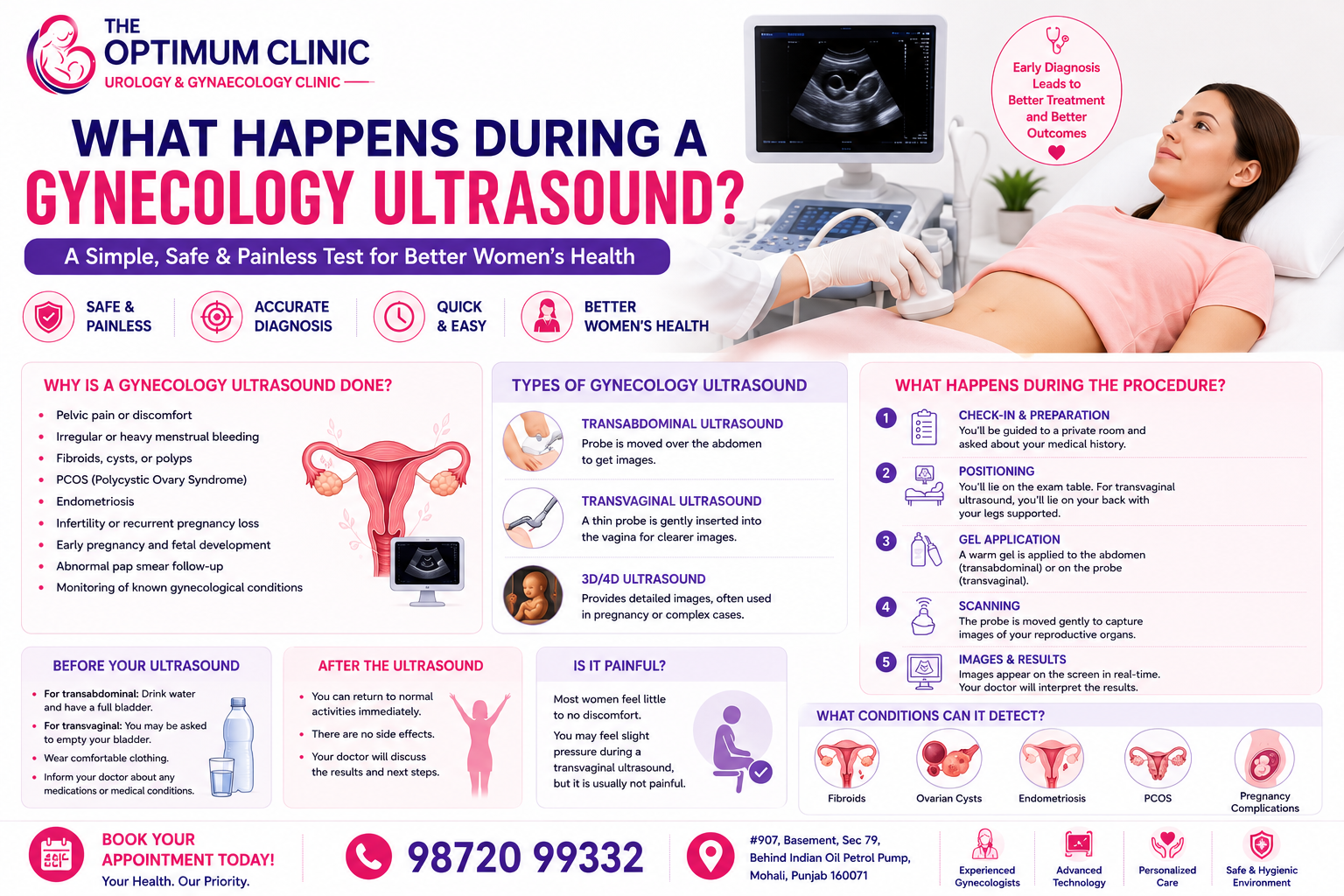
One of the most common questions women ask when they are advised to undergo an ultrasound is:
"Doctor, what exactly happens during a gynecology ultrasound? Is it painful?"
As a gynecologist, I understand that any medical test can feel intimidating when you don't know what to expect. The good news is that a gynecology ultrasound is one of the safest, quickest, and most informative diagnostic tests we use in women's healthcare.
Whether you are experiencing pelvic pain, irregular periods, heavy bleeding, fertility concerns, PCOS symptoms, or pregnancy-related issues, an ultrasound helps us understand what is happening inside your reproductive organs and guides us toward the most appropriate treatment plan.
In this guide, I will explain everything you need to know about gynecology ultrasounds, including why they are performed, how they work, what happens during the procedure, and what conditions they can help diagnose.
A gynecology ultrasound is a non-invasive imaging test that uses sound waves to create real-time pictures of a woman's reproductive organs.
The scan allows us to evaluate:
Unlike X-rays or CT scans, ultrasound does not use radiation, making it extremely safe for women of all ages, including pregnant women.
An ultrasound is often one of the first investigations recommended when a woman presents with symptoms affecting her reproductive health.
Common reasons include:
At our Gynecology Department, ultrasound plays an important role in diagnosing and monitoring many common women's health conditions.
This is the most commonly performed ultrasound and is done over the lower abdomen.
During the scan:
This method provides a broad overview of the pelvic organs.
A transvaginal ultrasound provides more detailed images of the uterus and ovaries.
During this procedure:
Most women experience only mild pressure and the procedure is generally well tolerated.
Used to:
This specialized scan is commonly used in fertility treatment and ovulation tracking.
Women undergoing fertility evaluation often benefit from this type of monitoring.
You may be advised to:
A full bladder improves visualization of the pelvic organs.
Before the scan begins, your doctor or sonographer will review your symptoms and medical history.
This helps ensure the ultrasound focuses on the areas most relevant to your concerns.
For a transabdominal ultrasound, you will lie comfortably on an examination table.
For a transvaginal ultrasound, you will lie on your back with your knees bent.
The ultrasound probe sends sound waves into the body.
These sound waves bounce off internal structures and create detailed images that appear instantly on the monitor.
During this stage, we carefully evaluate:
Several images are captured and stored for review.
These images help us create a complete assessment of your reproductive health.
Most gynecology ultrasounds take approximately 15–30 minutes.
You can usually return to normal activities immediately afterward.
This is perhaps the question I hear most often from patients.
The answer is reassuring:
If you feel discomfort at any point, you should inform the healthcare provider immediately.
Ultrasound can help identify ovarian changes commonly associated with PCOS.
Learn more about: PCOS Treatment in Mohali
Fibroids are one of the most common findings on gynecological ultrasound.
The scan helps determine:
Different types of ovarian cysts can be identified and monitored using ultrasound.
Although ultrasound cannot diagnose every case of endometriosis, it can reveal related findings that guide further evaluation.
Ultrasound is essential for monitoring healthy pregnancy development and identifying complications early.
I recommend seeking evaluation if you experience:
Early diagnosis often leads to better treatment outcomes and prevents complications.
As a gynecologist, I encourage women not to delay an ultrasound when it has been recommended. Many conditions such as fibroids, ovarian cysts, PCOS, and pregnancy-related complications can be identified early through ultrasound, allowing treatment to begin before symptoms become more serious.
Remember, an ultrasound is not something to fear. It is one of the safest and most valuable tools we have for understanding your reproductive health.
A gynecology ultrasound is a simple, safe, and highly effective diagnostic procedure that helps doctors evaluate the uterus, ovaries, and other reproductive organs. Whether you're dealing with menstrual problems, fertility concerns, pelvic pain, or pregnancy-related questions, ultrasound provides valuable information that supports accurate diagnosis and treatment.
If you have been advised to undergo an ultrasound, knowing what to expect can help you feel confident and prepared for your appointment.
The Optimum Clinic
#907 Basement, Sec 79, Behind Indian Oil Petrol Pump, Mohali
Call: 98720 99332